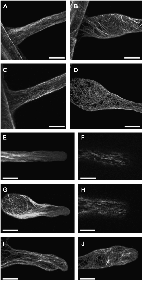
Figure 4.
Microtubule and F-actin organization is disrupted in living csld2-1 root hairs. A to D, F-actin (A and B) and microtubules (C and D) in the base of wild-type Col-0 (A and C) and csld2-1 (B and D). E to J, F-actin organization in the tip regions of wild-type Col-0 (E) and csld2-1 (G and I), and microtubule organization in the tip regions of wild-type Col-0 (F) and csld2-1 (H and J). F-actin and microtubules were visualized by GFP-ABD2-GFP and GFP-MBD2 fusions, respectively. Bars = 20 μm.

