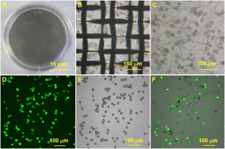
Figure 2.
Large-scale in vitro PG in liquid medium and pollen viability assay. A to C show the thin liquid layer method for PG and PTG. A, A 35-mm dish with a steel-wire net (80 μm) in it. The pollen grain suspension with 30 μL of basic medium was spread on the steel-wire net. (B) The pollen tubes under the steel-wire net after 4 h of incubation. (C) The collected pollen tubes after filtering through a 50-μm nylon mesh. D to F show FDA staining of the hydrated pollen grains after incubation in liquid medium for 45 min. D, FDA fluorescence image. E, Bright-field image. F, Merged image of D and E.

