
FIG. 3.
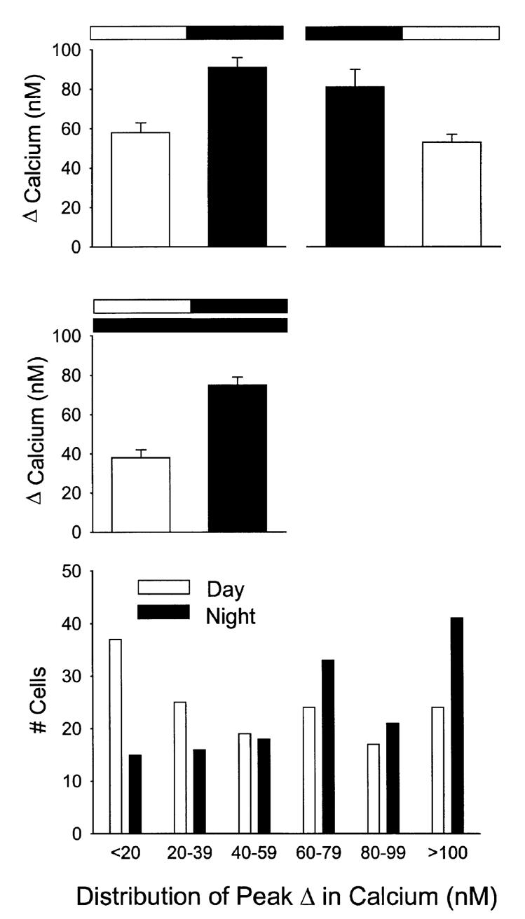
Diurnal rhythm in NMDA-evoked Ca2+ transients in SCN cells. In these experiments, NMDA-evoked Ca2+ transients were measured in SCN neurons in brain slices from animals during their day and compared to data obtained from brain slices from animals during their night. Animals were killed at either ZT 0 for the day group or ZT 12 for the night group. Each cell was sampled only once. Top left panel: NMDA-evoked Ca2+ transients peaked during night (day n = 83, night n = 102, P < 0.001). Top right panel: when the phase of the light-dark cycle to which the animals were exposed was reversed, so did the resulting rhythm (day n = 72; night n = 65, P < 0.05). Middle panel: in these experiments, animals were maintained in constant conditions and NMDA-evoked Ca2+ transients were measured in SCN neurons in brain slices from animals during their subjective day and compared to data obtained from brain slices from animals during their subjective night. Once again, NMDA-evoked Ca2+ transients peaked during subjective night (day n = 64; night n = 89, P < 0.001). Bottom panel: histogram illustrates the daily variation in the distribution of the NMDA-induced Ca2+responses. As has been previously reported (Colwell, 2000), in the presence of the ion channel blockers (TTX, D600), there was no significant day/night variation in the resting Ca2+ levels (day 89 ± 2 nM, n = 83; night 83 ± 1 nM, n = 102).