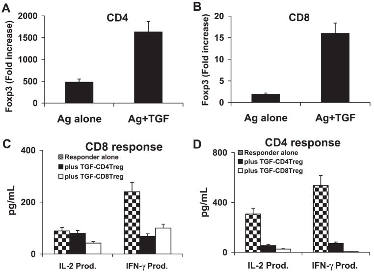
Figure 3.
TGF-β1 activated CD8 and CD4 suppressor T cells. (A, B) Responder CD4 (A) or CD8 (B) T cells were separated from IRBP1-20-immunized B6 mice by flow cy-tometry and were stimulated in vitro with antigen (10 μg/mL) and APCs in the absence or presence of 1 ng/mL of TGF-β1 for 3 days. The T cells were then separated by single-density gradient centrifugation, and cultured in IL-2 (10 ng/mL) medium for another 24 hours before the real-time PCR assessment of Foxp3 expression. The x-fold increase was then calculated (P < 0.01). (C, D) After the 3-day in vitro stimulation with antigen and TGF-β1, the CD8 (3C) or CD4 (D) T cells were assessed for suppressor activity of CD8 (C) and CD4 (D) T-cell responses. The responder T cells were freshly prepared CD8+ CD122− or CD4+ CD25− T cells from immunized B6 mice. After coculture of responder (4 × 10⁶ cells/well) and suppressor T cells (ratio 4:1) for 48 hours in 12-well plates with immunizing peptide (10 μg/mL) and APCs, the culture supernatants were sampled for measurement of IL-2 and IFN-γ by ELISA. The result shown is representative of those obtained in more than five experiments. TGF-CD4 Treg: TGFβ1-induced regulatory CD4 cells; TGF-CD8 Treg: TGFβ1-induced regulatory CD8 cells (P < 0.01).

