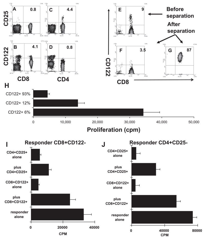
Figure 4.
Suppressor effect of the CD8+CD122+ T cell subset. (A–D) Unfractionated T cells from IRBP1-20-immuned mice were double-stained with PE-conjugated anti-CD25 or anti-CD122 antibody and FITC-conjugated anti-CD4 or anti-CD8 antibody. Some of the CD8 T cells express CD122, but not CD25, whereas some of the CD4 T cells express CD25, but not CD122. (E–G) Separation of CD8+CD122+ T cells from CD8+ CD122− T cells on a magnetic column (auto-MACS; Miltenyi Biotec GmbH, Bergisch Gladbach, Germany). (H) Unfractionated CD8 T cells (12% CD122+), CD122+-enriched CD8 T cells (93%), and CD8 T cells partially depleted of CD122+ cells (6% CD122+) were stimulated with 10 μg/mL of IRBP 1-20 in the presence of APCs and assessed for thymidine incorporation. CD8 IRBP-specific T cells showed increased proliferation when the CD122+ subset was partially depleted (P < 0.01). (I–J) Isolated CD8+CD122+ and CD4+ CD25+ T cells were suppressive of the CD4 (I) and CD8 (J) response. Magnetic bead-separated CD8+CD122− or CD4+ CD25− responder T cells were incubated for 48 hours in 96-well plates (4 × 10⁵ cells/well) with immunizing peptide (10 μg/mL) and APCs in the absence or presence of the indicated freshly prepared CD8+CD122+ or CD4+CD25+ Treg cells (1 × 10⁵ cells/well), and [3H] thymidine incorporation during the last 8 hours was assessed (P < 0.01).

