
Figure 1.
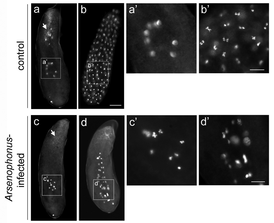
Early embryos from virgin Arsenophonus-infected females exhibit abnormal nuclear divisions. (a) A precortical embryo and (b) a cortical embryo, both from control LabII females. White arrow in (a) indicates endocellular Wolbachia bacteria at the posterior region of the embryo that accumulate into foci around maternal centrosomes. (a’) A higher magnification of eight interphase nuclei of the precortical embryo shown in (a). (b’) A higher magnification of anaphase nuclei of the precortical embryo shown in (b). (c and d) Defective pre-cortical embryos from virgin LabII(INF) females. White arrow in (c) indicates Wolbachia in the same region as in (a) but which fail to form foci due to the absence of maternal centrosomes (see text for explanation). (c’) and (d’) are higher magnifications of nuclei in (c) and (d), respectively. In all panels DNA is shown in greyscale. Scale bar equals 20 µm in (b) and 30 µm in (b’) and (d’).