Fig. 5.
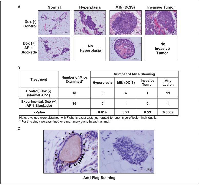
AP-1 blockade by Tam67 expression prevents premalignant lesions. A, normal-appearing mammary glands were collected and paraffin embedded from the trigenic mice that developed mammary tumors. Mammary tissue sections were stained with H&E. Selective fields containing hyperplasia, mammary intraepithelial neoplasia (MIN; DCIS), and invasive mammary tumors. B, comparison of the number of mice with premalignant lesions in doxycycline-treated or untreated trigenic mice as described in A. One normal-appearing number 3 mammary gland was removed from each mouse and the number and type of premalignant lesions in each mammary gland were assessed microscopically after H&E staining. C, representative fields of the mammary gland that showed DCIS from the mouse of the doxycycline-treated, AP-1-blocked experimental group. The mammary tissue was stained for TAM67 expression by anti-Flag immunohistochemical staining. Left, a normal duct that stained positive for TAM67 (arrows); right, the DCIS lesion that arose in this doxycycline-treated mouse. No TAM67 expression is seen in the DCIS cells.

