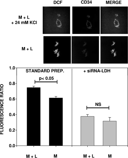
FIG. 5.
DCF fluorescence in CD34+ cell-lined channels within Matrigel. The top two rows of images show DCF fluorescence, as well as colocalization with the CD34+ cells in a Matrigel-lactide sample (M + L) with and without the topical addition of 24 mM KCl to cause cell depolarization. The bar graphs at the bottom show the ratios of DCF fluorescence (without versus with KCl) from different Matrigel samples (n = 3 different mice for each calculation). Where indicated, Matrigel samples were supplemented with siRNA specific to LDH (+siRNA-LDH). The fluorescence in the standard-preparation (standard prep.) Matrigel-lactide samples and Matrigel samples (M) was significantly greater than that in the siRNA samples.

