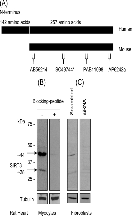
FIG. 1.
Characterization of antibodies recognizing two forms of SIRT3 from cardiac tissue. (A) Schematic representation of hSIRT3 and mSIRT3 with epitope map of different antibodies used in this study. The asterisk indicates an antibody that was used only for immunostaining of cells. (B) Western blot analysis with SIRT3 antibody (AP6242a) showing two specific bands of SIRT3 in cardiomyocytes, as determined by using a blocking peptide. (C) SIRT3 levels in rat heart fibroblasts were knocked down by using SIRT3-specfic siRNA. Cell lysate was analyzed by Western analysis with the same antibody as that in panel B. Note the reduced levels of both forms of SIRT3 after knockdown of endogenous SIRT3 levels.

