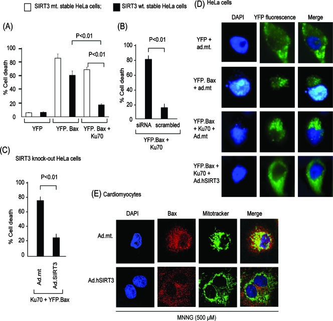
FIG. 11.
SIRT3 prevents Bax-mediated apoptosis by deacetylating Ku70. (A) HeLa cells stably expressing wt SIRT3 (black bars) or mt SIRT3 (white bars) were transfected with plasmids synthesizing YFP, YFP.Bax, or YFP.Bax and Ku70 together. The percentage of YFP-positive cells (yellow fluorescence) with apoptotic (fragmented) nuclei was scored 12 h after transfection. Values are the averages of three experiments; during each experiment >200 cells were scored. (B) HeLa cells stably expressing wt SIRT3 were given SIRT3-specific siRNA or scrambled RNA. We observed >80% reduction of the SIRT3 levels in cells to which siRNA was added, as shown in Fig. 4C. These cells were then transfected with plasmids encoding YFP.Bax and Ku70. Cell death was scored 12 h posttransfection. Values are the averages of four experiments. (C) Salvage of wt SIRT3 levels protects cells from Bax-mediated cell death. SIRT3-knockdown HeLa cells were infected with ad.hSIRT3 or the mt vector. After 12 h of virus infection, cells were transfected with YFP.Bax and Ku70 plasmids together. The percentage of YFP-positive cells with apoptotic nuclei was scored 12 h after transfection. Values are the means of four experiments with >200 cells scored in each plate. Nearly 80% of SIRT3 levels were recovered in these cells, as verified by Western blotting (not shown). (D) Representative picture of HeLa cells subjected to Bax-mediated apoptosis. Cells were infected with ad.hSIRT3 or the mt (ad.mt) vector. Twenty-four hours after viral infection, cells were transfected with different plasmids as indicated. The pictures of cells were taken 12 h posttransfection. Note the presence of fragmented apoptotic nuclei in cells transfected with YFP.Bax and YFP.Bax plus Ku70 (middle panels) but not in SIRT3-overexpressing cells (bottom panels). (E) SIRT3 blocks localization of Bax to mitochondria during genotoxic stress. Cardiomyocytes infected with ad.hSIRT3 or the mt (ad.mt) vectors were treated with MNNG for 6 h. Cells were stained with anti-Bax antibody (red). DAPI (blue) staining and Mitotracker (green) staining were utilized as markers of nuclei and mitochondria, respectively. Note shrunken nuclei in upper panels (ad.mt) depicting cell death. In these cells, Bax was totally merged with Mitotracker green (yellow in merged image) representing mitochondrial localization of Bax. In the lower panels, cells overexpressing wt SIRT3 and having well-preserved nuclei showed no Bax translocation to mitochondria (no yellow in merged image).

