FIG. 3.
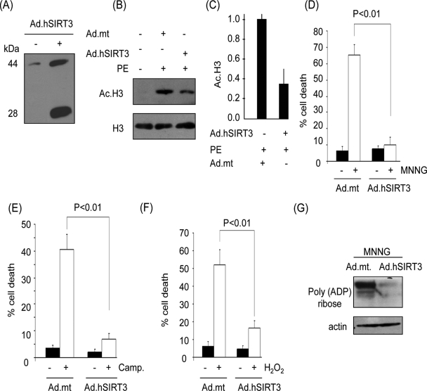
SIRT3 overexpression protects cardiomyocytes from genotoxic and oxidative stress-mediated cell death. (A) Expression levels of hSIRT3 in cardiomyocytes infected with adenovirus (Ad) vector for 24 h were determined by Western analysis with SIRT3 antibody PAB-11098. Note the presence of both forms of hSIRT3 in adenovirus-infected cells. (B) Deacetylase activity of hSIRT3 in cardiomyocytes. Cells infected with viral vectors, synthesizing the wt SIRT3 or an mt (benign) protein, were treated with PE to induce cellular acetylation. Acetylation of histones was determined by Western analysis. (C) Quantification of H3 deacetylation in SIRT3-overexpressing cells. (D to F) Cardiomyocytes overexpressing hSIRT3 or the mt protein were treated with MNNG (500 μM), camptothecin (Camp.; 10 μM), or H2O2 (200 μM). Cell death was determined 6 h after treatment by Hoechst and PI staining followed by FACS analysis. (G) Western analysis of the cell lysate with a poly(ADP-ribose) antibody, indicating PARP1 activity. Quantitative values are means ± standard errors of five to seven plates of three separate experiments.

