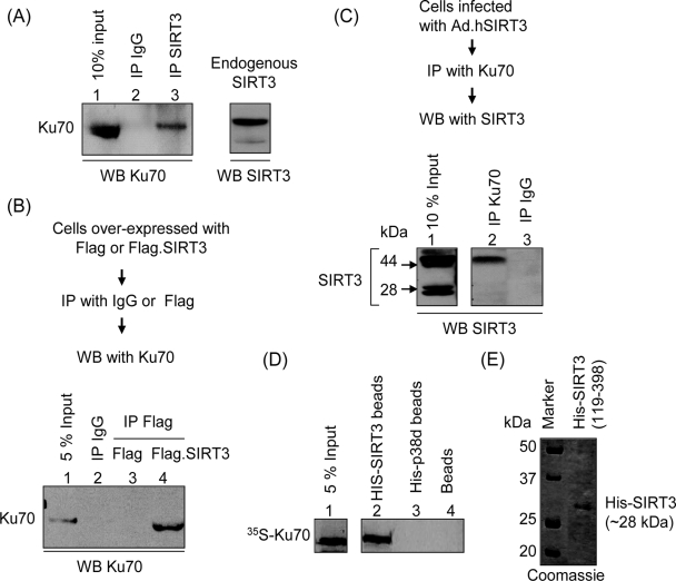
FIG. 8.
SIRT3 interacts with Ku70 in vitro and in vivo. (A) Endogenous SIRT3 interacts with Ku70. Cardiomyocyte lysate was subjected to IP with either nonspecific IgG or specific anti-SIRT3 antibody (AP6242a). Resulting beads were analyzed by Western blotting (WB) with anti-Ku70 antibody. (B) Cos7 cells were induced to overexpress with the Flag tag or the Flag-tagged SIRT3. Cell lysate was subjected to IP with either nonspecific IgG-conjugated beads or anti-Flag M2 agarose beads. Precipitated beads were analyzed by Western blotting with anti-Ku70 antibody. (C) Cells infected with ad.hSIRT3 vector were subjected to IP with Ku70 antibody, and the resulting beads were analyzed by Western blotting with anti-SIRT3 antibody (PAB11098). Note the presence of both forms of SIRT3 in the input lane, but only the long form of SIRT3 was pulled down by Ku70 in this assay. (D) In vitro protein binding assay. In vitro-synthesized [35S]methionine-labeled Ku70 was incubated with beads containing His-tagged 28-kDa SIRT3, His-tagged p38d, or nickel beads alone. His-tagged proteins were precipitated as nickel resin beads, and bound proteins were analyzed by SDS-PAGE. (E) Picture of a Coomassie blue-stained gel showing synthesis of the His-tagged 28-kDa form of SIRT3 from plasmid (His-SIRT3119-398).

