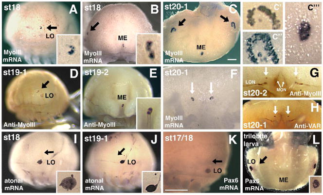
Figure 7. Limulus Pax6 and atonal are not expressed in developing eyes.
A–C,F) Myosin III mRNA expression in the rudimentary lateral eye (black arrows in A–C), early lateral ommatidial eyes (C″, C‴ show first ommatidial photoreceptors surrounded by crescent of rudimentary photoreceptors), rudimentary median eye (ME; B, C, C‴) and rudimentary ventral photoreceptors (arrows in F). The lateral sense organ (LO) is not involved in vision and does not express MyoIII mRNA (A and data not shown). D,E) Anti-Myo III antibody labels the embryonic rudimentary lateral eye (arrow in D), median eye (E) and more weakly the LO (D). F) High magnification of ventral photoreceptors (arrows) expressing MyoIII mRNA. G–H) Anti-MyoIII (G) and Anti-Visual Arrestin (VAR) staining of ventral photoreceptors (arrows) at approximately the same age as in F. These antibodies also label forming optic nerves innervating the brain from the rudimentary lateral (LON) and median (MON) eyes. I–J). Lp Atonal expression in the LO but not rudimentary lateral eye (black arrows point to eye). Insets show higher magnification of LO. K–L) Throughout ages when MyoIII mRNA and protein are expressed in rudimentary eyes or forming lateral ommatidial eyes, Pax6 mRNA is observed in the LO but is absent from these eyes (arrow points to rudimentary lateral eye). Panels A,D,I,J and K are lateral, B and E frontal, C and L are top-down dorsal, and F–H are ventral views. Scale bars in A, C = 500 μm, inset magnification is an additional 400X.

