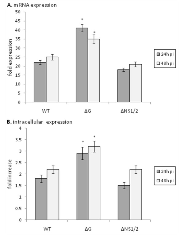
Figure 4.
ISG15 expression is increased in the absence of G protein expression. MLE-15 cells were mock-infected or infected with WT, ΔG, or ΔNS1/2 virus at a multiplicity of infection (MOI) of 1 for 24 h or 48 h as indicated. ISG15 message expression was measured by real-time PCR (A). Transcript levels were normalized to hypoxanthine guanine phosphoribosyl transferase (HPRT) expression and calibrated to the mock condition. (B) RSV stimulation of ISG15 protein expression was determined in MLE-15 cells that were mock-infected or infected with WT, ΔG, or ΔNS1/2 virus at a multiplicity of infection (MOI) of 1 for 24 h or 48 h as indicated. Cells were harvested and ISG15 levels determined by flow cytometry. Data is presented as fold-differences in protein expression relative to mock-infected cells. Differences in fold expression between virus infection groups were evaluated by Mann-Whitney U test and noted as significant as denoted by an asterisk. Data are shown as means ± standard errors (SE) of the means.

