Figure 4.

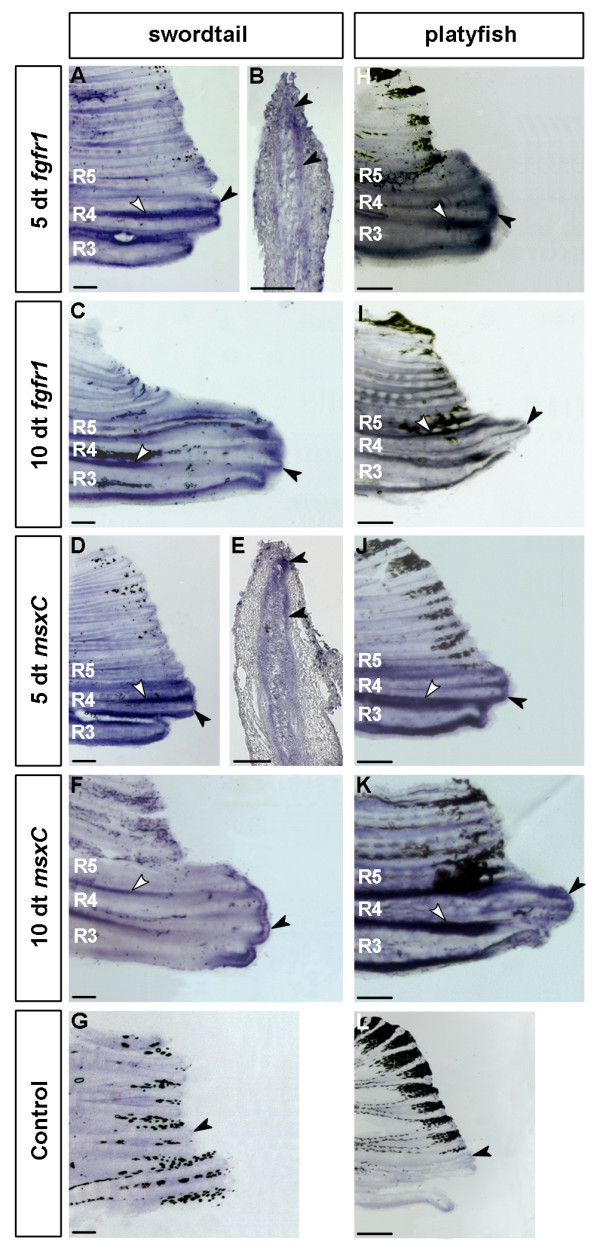
Expression of fgfr1 and msxC in the developing gonopodia of X. helleri and X. maculatus. fgfr1 and msxC are both expressed in developing gonopodia of X. helleri and X. maculatus. In X. helleri fgfr1 is up-regulated at 5 days (A) and 10 days (C) of treatment in mesenchymal cells (B) of the main gonopodium-forming rays 3–5 compared to control fins (G). In addition fgfr1 is strongly expressed in the interray tissue of those rays (A, C). As in developing swords, fgfr1 expression overlaps with msxC expression (D-F). In early stages of gonopodium development (5 dt) of the platyfish X. maculatus, the expression patterns of fgfr1 (H) and msxC (J) resemble that of X. helleri. Both genes are up-regulated in the same set of fin rays compared to untreated controls (L). Expression of both genes (I, K) at 10 dt is comparable to that of X. helleri with species-specific differences in the shape of growing rays. Black arrowheads indicate the expression in the distal part of the fin rays, white arrowheads indicate inter-ray expression. (X. helleri: n = 10 for every stage and probe; X. maculatus: 5 dt: n = 5; 10 dt and controls: n = 3; scale bars: A, C, D, F-L: 200 μm; B and E: 100 μm).
