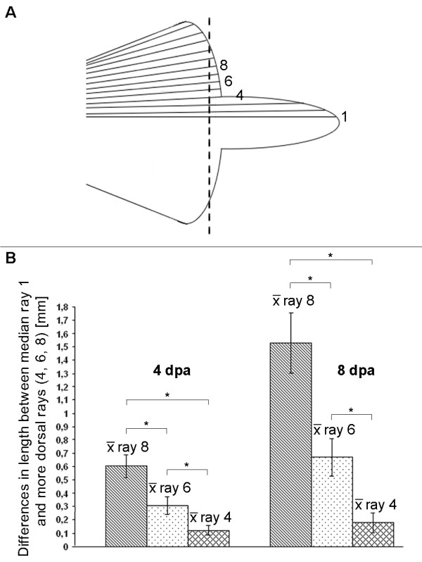
Figure 8.

Different regeneration rate of brushtail fin rays, depending on their position in the caudal fin. The regenerate's length of four different fin rays, highlighted in the schematic drawing of an adult brushtail fin (A) were measured at 4 days post amputation (dpa) and 8 dpa. The regenerate's length of the dorsal fin rays 4, 6 and 8 were then compared to that of the median fin ray 1. Dorsal fin rays regenerate more slowly than the median fin ray 1, shown as average length difference between fin ray regenerates (B). The difference in regeneration rate increases the closer a fin ray is located to the dorsal edge of the fin. The position dependence of regeneration rates is more obvious at 8 dpa (n = 11; *P < 0.00001, t-test).
