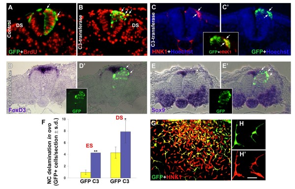
Figure 4.
Inhibition of Rho activity by C3-DNA stimulates neural crest (NC) delamination in ovo. (A-E) Transverse sections of embryos that received control green fluorescent protein (GFP) (A) or C3-DNA/GFP (B-E). Most delaminating cells contain bromo-deoxyuridine (BrdU)+ nuclei (arrows in (A,B)). (C,C' and inset) Electroporation of C3/GFP followed by HNK-1 labeling. (D,D'and inset) Electroporation of C3/GFP followed by in situ hybridization with FoxD3. (E,E'and inset) Electroporation of C3/GFP followed by in situ hybridization with Sox9. Note that delaminating, C3-containing cells (green) co-express all NC markers (arrows). Note also in (E,E') that the front of ventrally migrating precursors (arrowhead) is C3/GFP+ but has downregulated Sox9, which is normally transcribed in the dorsal neural tube and rapidly lost from emigrating NC. (F) Quantification of NC delamination at epithelial somite (ES) and dissociating somite (DS) levels. Electroporation of C3-transferase into hemi-NTs enhanced NC delamination opposite both epithelial somite and dissociating somite levels (*p < 0.05, **p < 0.01 compared to GFP-control). (G,H,H') C3-transfected cells co-express HNK-1. Neural primordia electroporated with C3/GFP were explanted. Delaminating NC cells co-express GFP and HNK-1 and exhibit a characteristic change in morphology. Bar: 42 μM (A-E); 50 μM (G); 20 μM (H,H').

