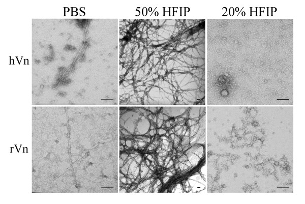
Figure 2.
Human vitronectin forms amyloid fibrils and oligomers. Left, Plasma-purified (hVn) and recombinant human vitronectin (rVn) aged in phosphate-buffered saline form a heterogeneous mixture of spherical and fibrillar structures as seen under TEM. Middle, Vitronectin treated with 50% HFIP promotes the formation of typical amyloid fibrils that are approximately 3–8 nm in diameter. Right, Incubation in 20% HFIP and 1 mM HCl enriches the population of spherical and protofibrillar oligomers, which range from 6–35 nm in diameter. Scale bar = 100 nm.

