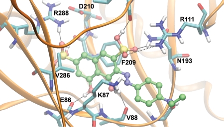
Fig. 5.
Predicted binding mode of S5. The most populated and lowest energy pose is shown for S5 docked into the TbREL1 crystal structure. S5 is shown in ball and stick, with carbons in green, nitrogens in blue, sulfur in yellow, oxygen in red, and hydrogens in white. Hydrogen bonds and salt-bridge interactions are shown with black lines. The protein residues are shown in licorice, with the same coloring except for carbons, which are cyan.

