Fig. 2.
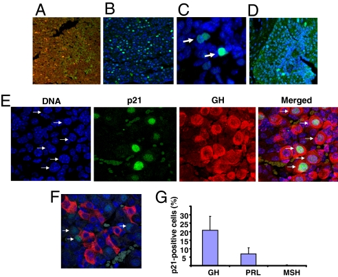
Pituitary p21 expression. Fluorescence immunohistochemistry of p21 expression in WT (A) and Pttg−/− (B) pituitary glands. High resolution (×100) confocal image shows intranuclear p21 expression in Pttg−/− pituitary anterior (C) and intermediate (D) lobes. Paraffin slides were labeled with p21 antibody (green). Here and below slides were counterstained with DNA-specific dye ToPro3 (blue). (E) Pttg deletion evokes aneuploidy and enhances p21 expression in GH-producing pituitary cells. Confocal image of Pttg-null pituitary tissue labeled with p21 antibody (green) and GH- antibody (red). (F) The same as E but stained with ACTH antibody (red). In E and F, arrows indicate aneuploid nuclei expressing p21. (G) Percent of hormone-producing cells coexpressing intra-nuclear p21 in Pttg−/− glands.

