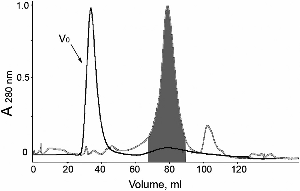
Figure 3.
The elution profile of Metridia luciferases purified from E. coli (black line) and insect Sf9 (gray line) cells on a HiLoad Superdex 75 column. The area highlighted in the dark gray color shows the collected fractions containing the active monomers of MLuc164. V0, void volume of the column.

