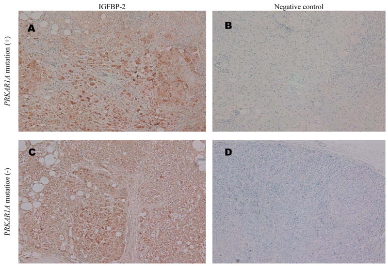
Fig. 2. Representative IGFBP-2 immunohistochemistry in PPNAD.

(A) PRKAR1A mutation-positive PPNAD section labeled with 1:50 rabbit anti-human IGFBP-2 antibody and visualized by the Envision Plus method using DAB color substrate and Harris’ haematoxylin counter-staining (original magnification X100).
(B) Adjacent section to that in Panel A, simultaneously processed the same way except using rabbit anti-human β amyloid precursor protein antibody as negative control primary antibody (original magnification X100).
(C) and (D) PRKAR1A mutation-negative PPNAD sections processed the same as (A) and (B), respectively (both original magnification X100).
