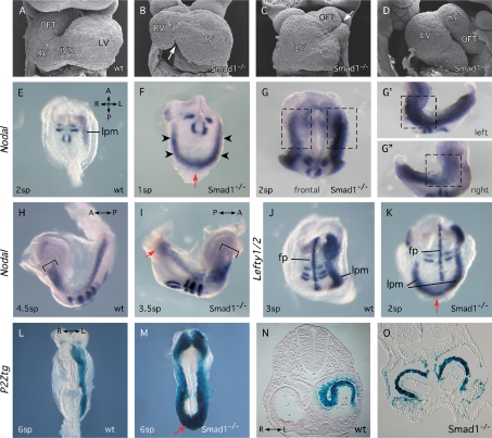
Figure 1.
Smad1-null embryos display LR defects. (A–D) SEM micrographs of hearts showing frontal (A–C) and lateral (D) views of wild-type (wt) (A) and Smad1−/− (B–D) embryos at 8.5–9.0 dpc. Arrows highlight abnormal interventricular sulcus (B) and bifid OFT (C). Hearts in C and D shows reversed and forward loops, respectively. (E–K) Whole-mount in situ hybridization showing expression of Nodal or Lefty1/2 in Smad1−/− mutant and wild-type embryos. Uncx4.1 signal indicates number of formed somite pairs (sp). Arrowheads (F) indicate robust precocious bilateral expression of Nodal at 1 sp. Red arrows (F,I,K) highlight expression of Nodal or Lefty2 across the caudal midline. Boxes in (G,G′,G″) highlight differences in Nodal expression levels in the anterior of Smad1−/− embryos. Brackets (H,I) indicate equivalent cranial extents of Nodal expression in wild type and Smad1−/− mutants. (L–O) LacZ staining in P2Ztg and P2Ztg;Smad1−/− embryos at 8.5 dpc highlighting molecular left isomerism in Smad1−/− mutants. (fp) Floorplate; (IVS) interventricular sulcus; (LV) left ventricle; (lpm) lateral plate mesoderm; (OFT) outflow tract; (RV) right ventricle.

