Figure 3.
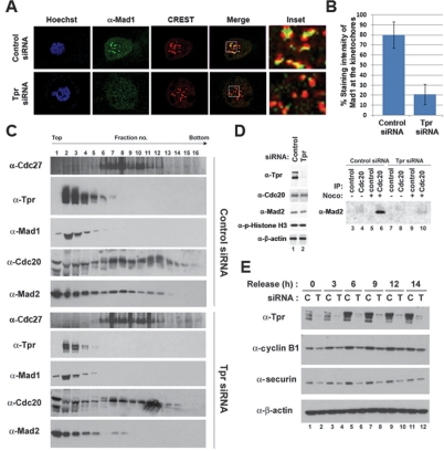
Tpr is important for activating Mad1 and Mad2. (A) Immunofluorescence analysis. HeLa cells transfected with siRNA were arrested in mitosis by nocodazole for 6 h and stained with indicated antibodies. (B) The staining intensity of Mad1 at kinetochores (n = 300) was quantified from control or Tpr-depleted cells (n = 50, each) selected at random in prometaphase stage. (C,D) HeLa cells transfected with siRNA were arrested in mitosis by nocodazole followed by mitotic shake-off to allow collection. (C) Whole-cell extracts were prepared and resolved on sucrose density gradient by centrifugation. Gradients were separated into 16 fractions and precipitated with TCA. The indicated protein was identified with immunoblot analysis. (D) Immunoprecipitation analysis. (Lanes 3–10) Equal amounts of HeLa cell lysates were subjected to immunoprecipitation with anti-Cdc20 antibody, and bound Mad2 was determined by anti-Mad2 antibody. (E) HeLa cells transfected with siRNA were synchronized at the G1/S boundary by thymidine double block. Cells were harvested at the indicated times after release from the G1/S boundary and subjected to immunoblot analysis.

