Fig. 4.
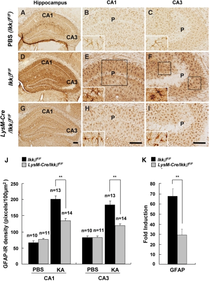
KA-induced astrocyte activation is reduced in LysM-Cre/IkkβF/F mice. Wild-type (IkkβF/F) (A–F) and LysM-Cre/IkkβF/F (G–I) mice were i.c.v. injected with either PBS (A–C) or KA (D–I). After 3 days, mice were sacrificed and cryosections were immunostained with anti-GFAP (A–I) antibodies. P: pyramidal cell layer. Scale bars: 100 µm. (J) Expression levels of GFAP in the CA1 and CA3 subfields of wild-type and LysM-Cre/IkkβF/F mice were quantified. (K) The mRNA levels of GFAP in the ipsilateral hippocampus were examined 36 h after KA injection. Data are presented as mean ± SEM. (Student's t-test, *P < 0.05, **P < 0.01; versus wild-type mice.)

