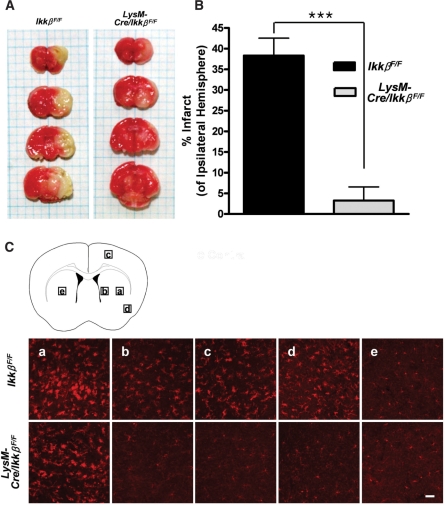
Fig. 9.
Infarct size and microglial activation following MCAO are reduced in LysM-Cre/IkkβF/F mice. Wild-type and LysM-Cre/IkkβF/F mice were subjected to transient MCAO for 1 h and reperfused. (A) After 71 h, the brains were removed, cut into 2-mm thick blocks and stained with triphenyl tetrazolium chloride. (B) The infarct area was measured and expressed as the percentage of the ipsilateral hemisphere. Data are presented as mean ± SEM. (***P < 0.001 by Student's t-test; versus wild-type mice; n = 4). (C) Cryosections of the second blocks were stained with anti-Iba-1 antibody. Representative images of five different regions were captured and are presented (ipsilateral: a–d, contralateral: e). Scale bars: 50 μm.

