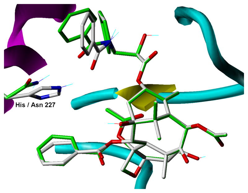
Fig. 3.
Binding model comparison for paclitaxel binding to brain β-tubulin and the corresponding H227N mutant. For the wild type complex, carbon atoms in the ligand and in His227 are rendered in grey, while for the mutation the corresponding carbons are green. All other atoms are rendered according to standard CPK coloring. Other receptor features are indicated according to underlying secondary structure as follows: magenta helices, yellow sheets and cyan coils.

