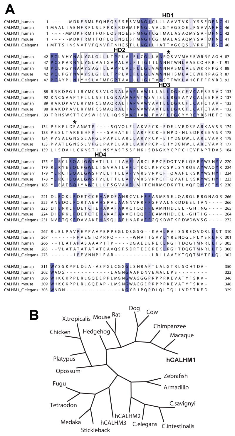
Figure 1. Alignment and phylogeny of CALHM1.
(A) Sequence alignment of human CALHM3, CALHM2, and CALHM1, and of murine and C. elegans CALHM1. Conserved sequences are highlighted in blue and sequence conservation is mapped in a color gradient, the darkest color representing sequences with absolute identity and lighter colors representing sequences with weaker conservation. Boxes denote hydrophobic domains 1–4 (HD1–4). Stars, predicted N-glycosylation sites on human CALHM1.
(B) Phylogenetic tree including human CALHM1 (hCALHM1).

