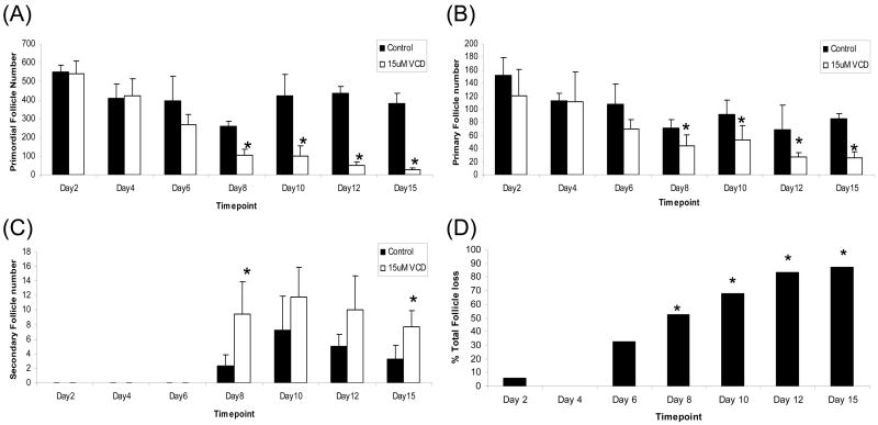
Figure 1. Time-course of VCD-induced ovotoxicity in B6C3F1 PND4 mouse ovaries.
Ovaries from PND4 B6C3F1 mice were cultured with control medium or 15μM VCD for 2, 4, 6, 8, 10, 12 or 15d. Following incubation, ovaries were collected and processed for histological evaluation as described in materials and methods. (A) Primordial, (B) Primary, and (C) Secondary follicles were classified and counted. (D) Total follicle loss in VCD treated ovaries as a percentage of control treatment. Values are mean ± SE total follicles counted/ovary, n=5; * Different (p<0.05) from control. NOTE: Secondary follicles do not form until day 8 in culture.

