
Figure 4. Effect of VCD on mEH, GSTpi and GSTmu protein expression on day 8.
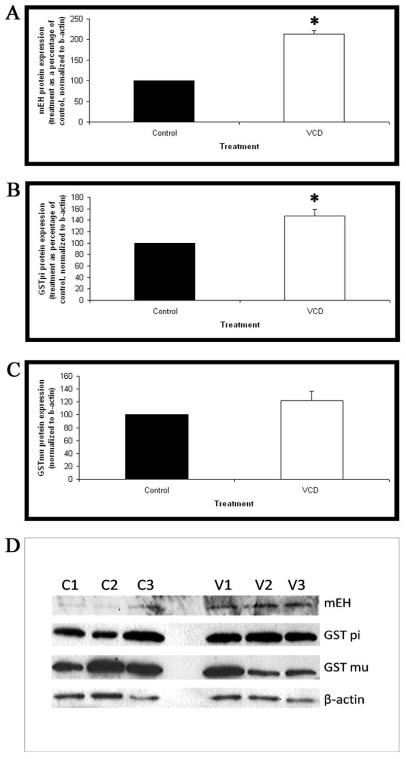
Ovaries from PND4 B6C3F1 mice were cultured with control medium or medium containing 15μM VCD for 8 days. Following incubation, total protein was isolated and Western blotting carried out for (A) mEH, (B) GST pi and (C) GST mu. (D) Representative western blots for mEH, GST pi, GST mu and beta-actin (Control=C1,C2,C3; VCD=V1,V2,V3). Values are normalized to beta-actin protein and expressed as treatment as a percentage of control mean ± S.E.; n=3. * Different (p<0.05) from control.