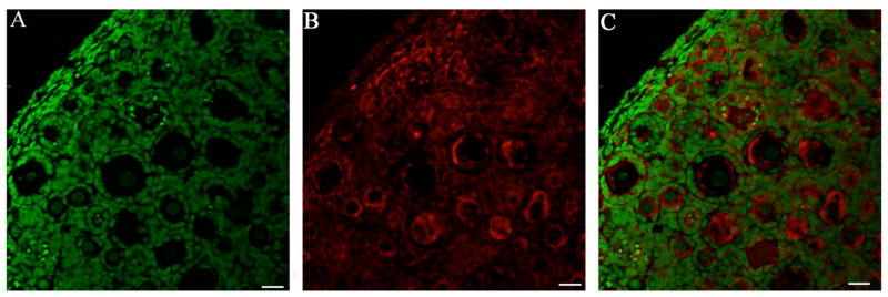
Figure 6. GST mu protein expression in PND4 cultured ovary.
B6C3F1 ovaries were cultured with control media for 8 days and processed for confocal microscopy as described in materials and methods. (A) Genomic DNA (green YOYO1 stain), (B) GST mu (Cy-5 red stain) and (C) Combined overlay of GST mu (Cy-5 red stain) and Genomic DNA (green YOYO1 stain) at 40X magnification. Scale-bar equal to 25μM.

