
Figure 5. POMx inhibition of JNK activation in mouse macrophages.
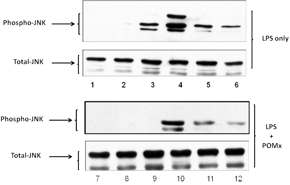
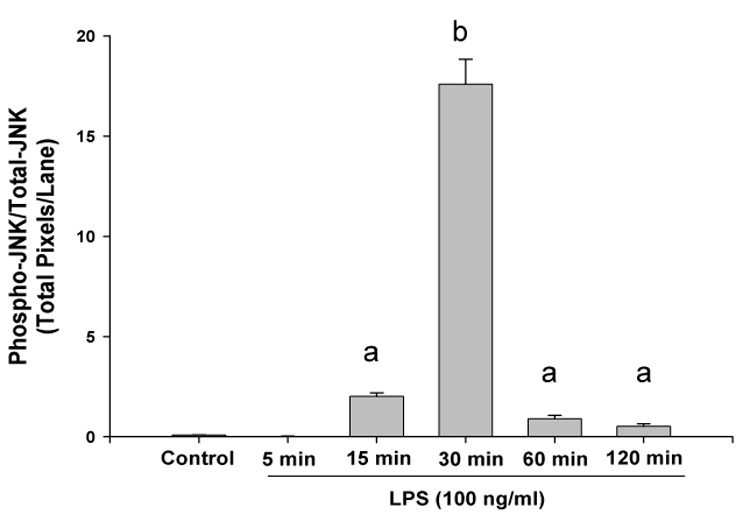
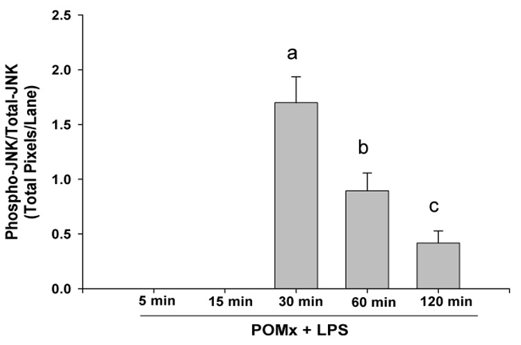
RAW 264.1 mouse macrophages were stimulated with LPS alone, or pre-treated with POMx and then stimulated with LPS for different time periods and cell lysates were prepared for immunoblotting. Immunoblots were probed with antibodies specific for JNK and phospho-JNK. (A) Lanes 1–6, control and cells stimulated with LPS for 5, 15, 30, 60 and 120 min; Lanes 7–12, control, cells pre-treated with 20 µg/ml POMx and then stimulated with LPS for 5, 15, 30, 60 and 120 min. (B & C) Phospho-JNK/Total JNK ratio for results shown in 5A.