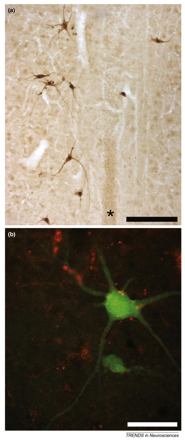
Figure 2.
Photomicrographs of longitudinal sections (40 microns) from the cervical spinal cord of an adult rat. Within 48 h following delivery of the transsynaptic tracer pseudorabies virus (PRV) to the left hemidiaphragm, the ipsilateral phrenic motoneuron pool becomes infected with the retrogradely transported virus. Subsequently (64 h; [a,b]), pre-phrenic interneurons become transsynaptically infected. (a) Antibodies against PRV were used to immunocytochemically visualize PRV-infected cells at around laminae VII/X. These interneurons are bilaterally distributed around the central canal and spinal cord midline (*). (b) This pre-phrenic interneuron (green) has been retrogradely infected with the PRV that results in expression of green fluorescent protein. Projections from the ventral respiratory column in this animal were anterogradely labeled using MiniRuby (red). These projections appear in close contact with the soma and dendrites of the interneuron, which could suggest synaptic contact. This finding (described in detail by Lane et al. [85]) indicates that whereas there is substantial monosynaptic bulbospinal-phrenic projection, there is also anatomical evidence for a polysynaptic pathway in the phrenic circuit of the rat. This pathway might serve as an alternative to direct monosynaptic projections from the medulla following spinal cord injury and represent a viable therapeutic target. Scale bar is 200 (a) and 50 (b) micrometers.

