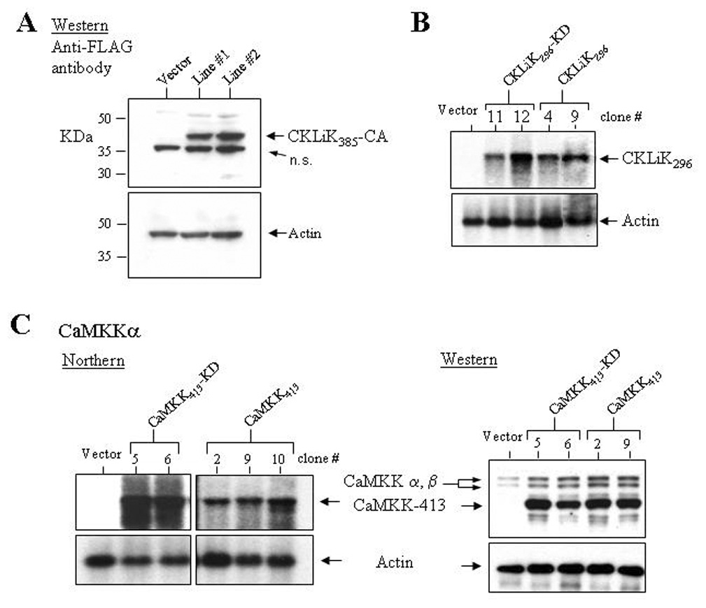
Figure 3. Ectopic expression of mutant CKLiK and CaMKKα in EML cell lines.

(A) A Western blot containing protein lysates from two independently generated EML cell lines stably transduced with CKLiK385-CA or an empty vector was probed with an anti-FLAG antibody. Below is shown the same blot probed with an anti-actin antibody. (B) Ectopic expression of CKLiK296 and CKLiK296-KD in clonal sublines of transduced EML cells was demonstrated by northern analyses. (C) Ectopic expression of constitutively active and kinase dead forms of CaMKKα (CaMKK413 and CaMKK413-KD, respectively) in EML cells was confirmed by northern assays and a Western blot probed with anti-CaMKK. Shown in the Western blot is expression of the endogenous CaMKKα and β isoforms, and the shorter, truncated forms. Levels of actin expression are shown below each blot.
