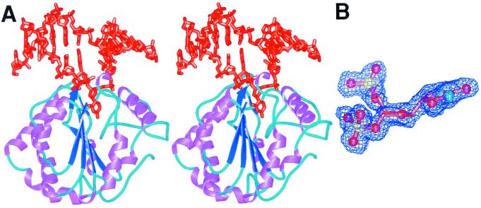
Figure 2.
Cocrystal structures of UDG bound to uncleaved substrate and cleaved product DNA. (A) dΨU-containing DNA (orange) binds UDG near the C-terminal end of its central β-sheet (dark blue arrows), which is surrounded by eight α-helices (purple). (B) Experimental electron density defines the stereochemical deformation and intact bond for the substrate dΨU complex. The glycosylic bond of the dΨU (orange carbon tubes, red oxygens, blue nitrogens, yellow phosphorus) is not cleaved, as demonstrated by the simulated-annealed omit map (blue) contoured at 2σ. The normally trigonal planar 1-position is clearly distorted out of the plane of the uracil ring toward a tetrahedral geometry. Difference maps of the active-site center are flat, indicating that the distortion is accurately depicted by the crystal structure.

