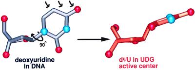
Figure 4.
Deoxyuridine (gray carbon tubes, red oxygens, blue nitrogens) in DNA is severely distorted by the UDG active center to achieve the observed conformation of the dΨU (orange carbon tubes, red oxygens, blue nitrogens). The left side of the large arrow is deoxyuridine in the conformation normally found in DNA. The arrow implies the observed flipping of the substrate nucleotide out of the DNA helix, which results in the altered position of the 5′P. When flipped into the UDG active center (right side of large arrow), the uracil ring is rotated ≈90° on its N1–C4 axis to a χ angle of 177°. Furthermore, the deoxyribose sugar of the enzyme bound substrate is flattened to a mild C3′-exo, which raises the uracil to a semiaxial position. The normally trigonal planar 1-position of uracil is strained to an almost tetrahedral geometry. The small arrows indicate the steric hindrance, which causes the deformation at the uracil 1-position. The conformation of deoxyuridine in DNA (gray) is derived from the conformation of deoxythymidine in a G/T mismatch (Protein Data Bank accession code 113D).

