
Figure 1. Comparison of NF-κB pathway between VDR(+/-) and VDR(-/-) MEFs.
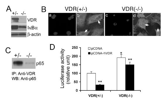
(A) Basal levels of IκBα protein in VDR(+/-) and VDR(-/-) cells determined by Western blotting. Also shown is the VDR levels determined using anti-VDR antibody. (B) Confocal microscopy showing increased p65 nuclear translocation in VDR(-/-) cells. VDR(+/-) and VDR(-/-) MEFs were stained with 4-diamidino-2-phenylindole (DAPI) to visualize the nucleus (a and c) or with anti-p65 antibody (b and d). (C) Protein-protein interaction between VDR and p65. VDR(+/-) and VDR(-/-) cell lysates were immunoprecipitated with anti-VDR antibody (IP), and then the precipitated complex was probed with anti-p65 antibody by Western blotting (WB). (D) Suppression of NF-κB transcriptional activity by hVDR over-expression. VDR(+/-) and VDR(-/-) MEFs were co-transfected with pNF-κB-Luc reporter, pRL-TK and pcDNA3.1 or pcDNA-hVDR, and luciferase activity was determined after 24 hours. *, P<0.05 vs. VDR(+/-) cells; **P<0.05 vs. corresponding pcDNA control.