
Figure 2. IL-6 synthesis in MEFs.
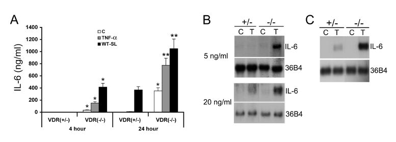
(A) IL-6 secretion. VDR(+/-) and VDR(-/-) MEFs were untreated (C) or treated with TNFα (20 ng/ml) or Salmonella (WT-SL) for one hour, and incubated in media containing Gentamicin for 4 or 24 hours. IL-6 secretion into the media was determined using a mouse IL-6 EIA kit. Note the basal IL-6 production in unstimulated VDR(+/-) cells was below the detectable limit. *P<0.05, **P<0.001 vs. corresponding VDR(+/-) value. (B and C) IL-6 mRNA expression. (B) VDR(+/-) and VDR(-/-) MEFs were untreated (C) or treated with 5 ng/ml (upper panel) or 20 ng/ml (lower panel) TNFα (T) for 6 hours. (C) VDR(+/-) and VDR(-/-) MEFs were untreated (C) or treated with 1 ng/ml IL-1β (T) for 6 hours. IL-6 mRNA levels were determined by Northern blotting. 36B4 is the internal loading control.