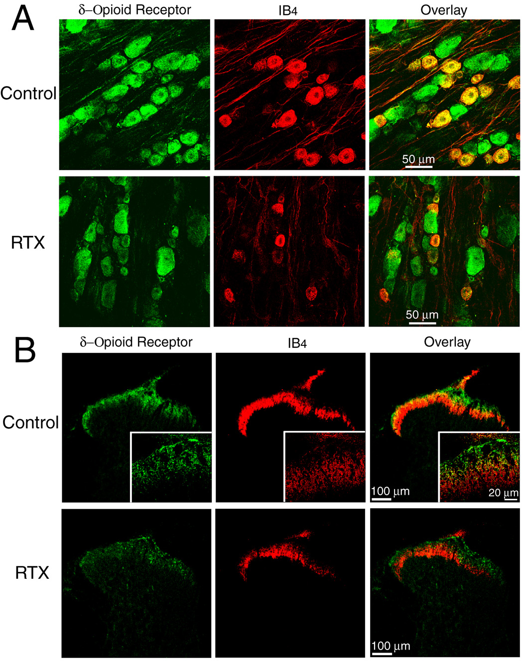
Fig. 2.
Confocal images showing the effect of RTX on δ-opioid receptor- and isolectin B4 (IB4)-positive dorsal root ganglion (DRG) neurons and afferent terminals in the spinal cord. A: representative confocal images showing δ-opioid receptor-immunoreactive (green) and IB4-positive (red) DRG neurons from one control and one RTX-treated rat. B: confocal images showing δ-opioid receptor-immunoreactive (green) and IB4-positive (red) afferent terminals in the spinal dorsal horn of one vehicle control and one RTX-treated rat. Colocalization of δ-opioid receptor-immunoreactivity and IB4-labeling is indicated in yellow when two images are digitally merged. All images are single confocal optical sections.

