
Figure 2. In vivo expression of YC3.6-AAV.
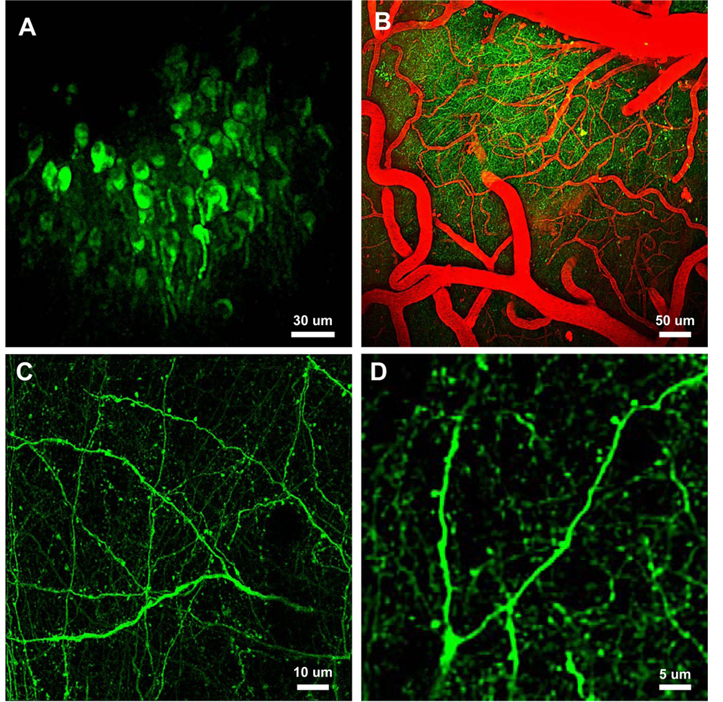
A–D) In vivo multiphoton images deep in the neocortex of adult mice. A) Cell bodies 300–550 microns below the brain surface. B) Full field images of YC3.6-filled neuritis and fluorescent angiogram (red). C) High-resolution images of axons, dendrites and spines in layer I of a wildtype mouse. D) Higher magnification images of a dendritic tree and corresponding spines. Detailed analysis of cellular and sub-cellular morphology was possible with YC3.6.