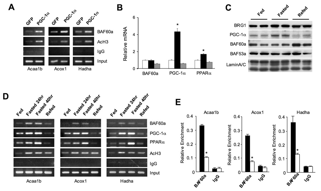
Figure 6. Recruitment of BAF60a to FAO Genes is Induced by Fasting.
(A) ChIP assay using BAF60a or AcH3 antibodies in H2.35 cells transduced with GFP or PGC-1α adenoviruses. PCR primers flanking PPRE present in the promoters of Acaa1b, Acox1, and Hadha were used.
(B) qPCR analysis of BAF60a, PGC-1α, and PPARα expression in fed (open), fasted (24 hrs, filled), and fasted/refed (24/20 hrs, grey) mouse livers. Data represent mean ± SEM, n=4. *p<0.05.
(C) Immunoblots of liver nuclear extracts using indicated antibodies.
(D) ChIP assays with chromatin extracts prepared from fed, fasted and refed mouse livers using indicated antibodies. PCR primers are the same as in (A).
(E) ChIP-qPCR assay with chromatin lysates prepared from 24-hr fasted wild type (filled) or PGC-1α null (open) mouse livers using BAF60a antibody. Data represent mean ± stdev, *p<0.01 wild type vs. PGC-1α null.

