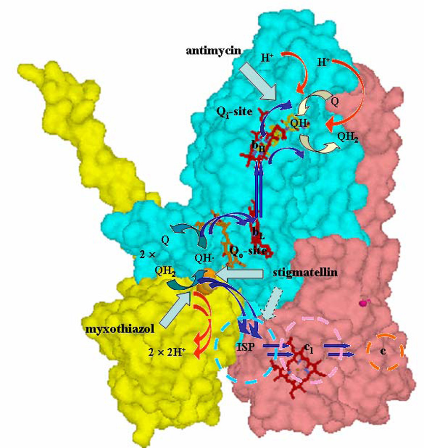
Fig. 1. The modified Q-cycle mechanism.

The mechanism demonstrated in the 1980’s, superimposed on the Rb. sphaeroides structure (PDB file 2QJK). Electron transfers are shown by blue arrows; H+ release or uptake by red arrows, exchange of Q or QH2 by open blue-green (Qo-site) or yellow (Qi-site) arrows; inhibition sites by cyan arrows. See text for description of operation.
