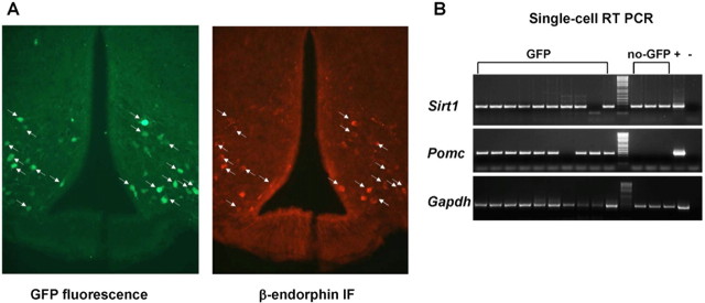
Figure 3.
POMC neurons express Sirt1 mRNA. A, Photomicrographs of brain slices from Pomc-GFP mice stained for β-endorphin. Green represents GFP fluorescence, and red represents β-endorphin immunofluorescence (IF). Arrows indicate neurons expressing both GFP and β-endorphin. B, Agarose-gel analysis of single-cell RT-PCR. Fluorescent (GFP) and POMC neighboring, nonfluorescent (no-GFP) cells were harvested from Pomc-GFP hypothalami. PCR products corresponding to SIRT1, POMC, and GAPDH were visualized. All pairs of primers were designed across at least one intron, allowing discrimination between genomic- and cDNA-specific amplicons. Hypothalamic cDNA was used as positive control (+), whereas the same PCR mix without template was used as negative control (−).

