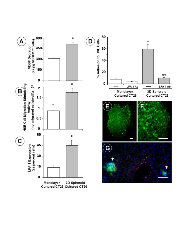
Figure 2.
(A) VEGF secretion by cultured CT26 cells. Supernatants were obtained on the 18th hour of incubation of CT26 cells, and the concentration of VEGF was determined by ELISA. (B) Hepatic sinusoidal endothelium (HSE) cell migration in response to conditioned media from CT26 cells. Primary cultured HSE cells were incubated for 48 hours with CT26 cell-conditioned media and endothelial cell migration was assayed across type-I collagen-coated inserts. (C) Flow cytometric study on LFA-1 expression. CT26 cells were incubated for 30 minutes at 4°C with 1 μg/106 cells of rat anti-mouse LFA-1 antibody followed by conjugated alexa-IgG2a anti-rat antibody labeling. (D) Adhesion assays of CT26 cells to HSE cells. CT26 cells received 1μg/ml anti-murine LFA-1 antibodies 30 min prior to the adhesion assay. All data from A-to-D studies represent average values ± SD from 3 different experiments (n = 18). Statistical significance: (*) p < 0.01 as compared to monolayer-cultured CT26 cancer cells; (**) p < 0.01 as compared to untreated CT26 cancer cells. (E-F) Inmunofluorescence pictures on LFA-1 expression (green staining) by 3D-spheroid-cultured CT26 cells and (G) a vascular hepatic micrometastases (arrows) on the 7th day after intrasplenic injection of monolayer-cultured CT26 cells. Red staining corresponds to ASMA-expressing fibroblasts around a terminal portal venule and some sinusoids. Scale bar: 20 μm.

