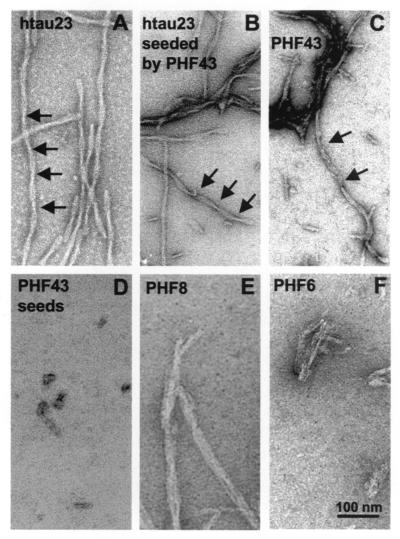
Figure 3.
Electron micrographs of fibers obtained from the self assembly of τ or τ peptides. Assembly conditions were the same as in Fig. 2 (20 μM protein, 5 μM heparin), except E and F (660 μM peptide, 660 μM heparin). A–C show mostly twisted filaments (width ≈10–20 nm) resembling Alzheimer PHFs polymerized from, (A) hτ23, (B) hτ23 plus seeds made from PHF43, and (C) PHF43. D shows the seeds obtained by sonication of PHF43 fibers. E and F show filamentous aggregates with variable diameters obtained from the short peptides PHF8 and PHF6.

