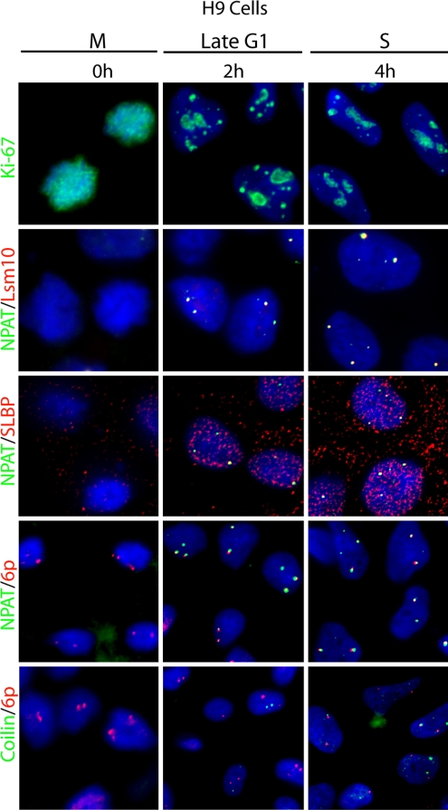
Fig. 3.
Association of p220NPAT and 3′-end processing factors with histone gene loci during the hES cell cycle. Mitotically synchronized hES cells at various cell cycle stages were monitored by IF microscopy for association of Lsm10 or SLBP (red) with p220NPAT (green; rows 2 and 3), and spatial linkage of the histone gene cluster at 6p22 (red) with p220NPAT or coilin (green; rows 4 and 5). Ki-67 (green) staining (row 1) was done to establish cell cycle position, and DAPI staining (all rows; blue) was used to visualize the nucleus.

