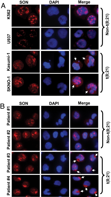
Fig. 6.
Abnormal cytoplasmic localization of SON in t(8;21)-positive leukemic cells. (A) Two leukemic cell lines without t(8;21), K562 and U937, and two t(8;21) leukemic cell lines, Kasumi- 1 and SKNO-1, were stained with SON antibody and DAPI (for DNA). (B) Blood cells from four different AML patients were stained with SON antibody and DAPI, and analyzed by fluorescence microscopy. Patient 1, FAB M2 subtype, non-t(8;21); patient 2, FAB M4 subtype, non-t(8;21); patients 3 and 4, FAB M2 subtype, t(8;21) -positive. Arrows indicate cytoplasm-localized SON. Pictures are of representative features of each cell line and patient samples from >1000 cells analyzed by fluorescence microscopy.

