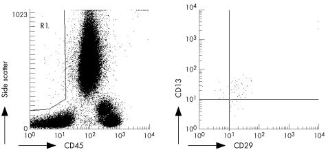
Figure 1 Identification of circulating mesenchymal cells (cMSCs) by flow cytometry. Peripheral blood samples were stained with monoclonal antibodies as described in the text. A gate containing medium‐high forward and side scatter non‐haematopoietic cells was drawn (R1, left). cMSCs were defined as positive for the markers CD29 and CD13, within region R1 (right).

An official website of the United States government
Here's how you know
Official websites use .gov
A
.gov website belongs to an official
government organization in the United States.
Secure .gov websites use HTTPS
A lock (
) or https:// means you've safely
connected to the .gov website. Share sensitive
information only on official, secure websites.
