Figure 1.
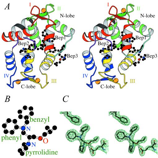
The overall structure of bepridil-3Ca2+-cTnC. (A) Stereo view of bepridil-3Ca2+-cTnC. cTnC comprises an N- and a C-terminal lobe. Each lobe consists of two helix–loop–helix EF-hand domains connected by a linker. The nomenclature is as follows: domain I (red) starts with helix A, followed by loop I, then helix B followed by the N-terminal linker (cyan); domain II (green) starts with helix C, followed by loop II, then helix D followed by the central linker (not included in the current structure because of disorder); domain III (yellow) starts with helix E, followed by loop III, then helix F followed by the C-terminal linker (purple and discontinuous at residues 125–126 because of disorder); domain IV (blue) starts with helix G, followed by loop IV, then helix H. There is also a helix N (black) N-terminal to domain I, which is present in TnC but not in CaM. There are two small antiparallel β-sheets in cTnC: one between loops I and II, and the other between loops III and IV. Three Ca2+ ions, bound to domains II, III, and IV, are shown as orange spheres. Three bepridil molecules (balls and sticks) are bound to cTnC: Bep1 mainly to the N-lobe, Bep2 mainly to the C-lobe, and Bep3 to both lobes. (B) Ball-and-stick representation of the calcium-sensitizing agent bepridil. (C) Electron density around Phe27 (upper left), Bep1 (center), and Glu96 (lower right) in a (2|Fo| − |Fc|) map contoured at 1.0σ. The positively charged pyrrolidine ring of Bep1 forms a salt bridge with the side chain of Glu96.

