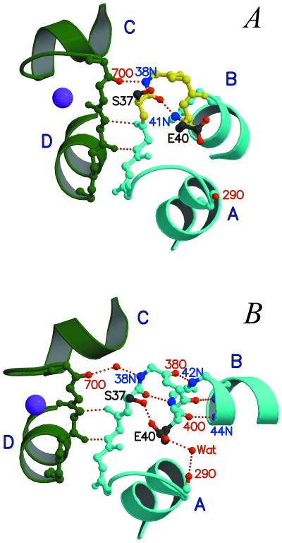
Figure 3.
A detailed view of the bepridil-induced opening of the N-lobe. (A) In 3Ca2+-cTnC (11), helix B displays a kink due to the nonhelical torsion angles of residues 37–40 (yellow). (B) The binding of bepridil results in a rearrangement of the hydrogen bonding pattern in the small β-sheet between loops I and II and in the N-terminal region of helix B. Consequently, helix B is extended at the N terminus to include residues 37–40 (cyan), and its C terminus is swung away from helix A. Note that the breaking of the 38N⋅⋅⋅⋅ 70O hydrogen bond also leads to a small decrease in the C–D interhelical angle. Bep1 is not shown.

